Monday, 22 February 2016

TRANSPORT IN PLANTS


1- Parenchyma 
- living cells
- round in shape
- thin cell walls
- large vacuole
- functional cell e.g; Chlorenchyma cell--> mesophyll cells
- large intercellular spaces


2- Collenchyma
- living cells
- rectangular in shape
- thick cell walls
- smaller vacuoles
- can be functional + supportive
- no intercellular spaces
- in bends of plant


3- Sclerenchyma
- dead cell
- present as long fibres
- very thick cell walls
- no intercellular spaces
- non-functional but supportive
- scattered specially around vesicular bundles


Transport of water: from soil to plant
Soil has higher water potential gradient as compared to root hair cells, water molecules enter root hair cells by osmosis down the concentration gradient
Root hair cells has a long extension which increase surface area for better absorption it has thin cell walls and a lot of mitochondrea


Q) What are the different pathway for transfer of water molecules across cortex cells?

1- Symplast pathway:
                                  osmosis as water molecules pass through cell membrane and cytoplasm, and are then taken up to the next cell down the water potential gradient

2- Vacular pathway:
                                 kind of symplast pathway when water passing through cytoplasm also enters the vacuole

3- Apoplast pathway:
                                In which water doesn't enter the cytoplasm but diffuses across the cellulose cell wall due to adhesion and cohesion


(Sucrose actively loaded into sieve tube elements decreases the water potential causing the hydrostatic pressure to increase)


Translocation: 
- transport of assimilates from source to sink (through phloem vessels)
- sucrose and amino acids assimilates exose

- Source: part of plant where assimilates are loaded up into the vessels  e.g; photosynthesising leaves or storage organs (green leaves)

Sink: part of plant where unloading takes place e.g; growing part of plant (storage roots)


Structure of Phloem~
* made from specialized cells called sieve elements which have cytoplasm, Organelles are absent
* they have sieve pores in the cross walls which results in formation of sieve plates
* they are accompanied by companion cells adjacent to them
* they are lots of plasmodesmata between sieve elements and companion cells
* companion cells are true cells with usual cytoplasm, a small vacuole and many mitochondrea since they perform active transport


(water movement from roots to leaves: evaporation of water from the messophyll cell walls)


Loading of Assimilates:
mesophyll cells photosynthesize to form glucose which can be stored as starch. rest of it becomes sucrose which is to be loaded into the sieve element
+ the adjacent companion cell with use of ATP pumps hydrogen ions into mesophyll cells by active transport
+ these hydrogen ions being in higher assimilation move back into companion cells through a co-transport prokin along with sucrose molecule against concentration gradient
+ this lowers the solute potential and water potential in companion cell
+ water enters companion cell from surrounding cells by osmosis
+ this increases the hydrostatic pressure and causes the sucrose solution to diffuse into sieve element


Passage of assimilates through sieve elements~
- hydrostatic pressure that develops in a sieve element (the source) because of osmosis decides for direction of movement as it goes down the pressure gradient, from higher pressure to lower pressure. this results in translocation from source to sink


Unloading of Assimilates:
In sink the sieve element having higher concentration of sucrose than the adjacent cells, results in diffusion of sucrose and amino acids down the concentration gradient
+ concentration in always kept lower due to enzyme invertase which converts sucrose into glucose and fructose


Transpiration~
evaporation of water molecules from cell walls of mesophyll cells which then escapes through stomata.

Stomata: loss of water vapour from leaves
                 stomata are open for gas exchange
                 stomata opens as light intensity increases


Factors of transpiration:

1- temperature increases, transpiration increases
2- wind increases, transpiration increases
3- humidity increases, transpiration decreases
4- light intensity increases, transpiration increases


*Protometer is used to measure transpiration rate*


Xerophytes:
- desert
- hot climate
- lesser water supply


1) waxy cuticle
2) sunken stomata
3) trichomes (hair)
4) surface area decreases
5) fleshy green
6) very deep and wide spread
7) less frequent stomata per unit area


Hydrophytes:
- stomata on uper epidermis
- are spaces (large)


Mesophytes:
- normal (leaves) plants


Thursday, 18 February 2016

CELL AND NUCLEAR DIVISION

            Structure of DNA

- DNA is a poly-nucleotide
- DNA is in form of a double helix in which 2 strands are coiled with each other


Nucleotide: it is made from a combination of deoxyribose sugar (pentose) phosphate molecule and a nitrate base


*In 1 strand many nucleotides are bound with each other through 3-5 phosphate bonds,

*strand opposite to first 1 is called anti parallel since it has 5-3 linkage

*the 2 strands are complementary to each other in such a way that adenine is always with thymine and cytosine is always with guanine

*complementary base attached to each other by hydrogen bonds

*there are 2 bonds between adenine and thymine. 3 between cytosine and guanine

*among the 4 nucleotides 2 are purine ans rest are pyrimidine


Complementary base paring
1- purine and pyrimidine are different sizes
2- adenine forms the same number of H+ bonds with thymine ans uracil
3- the base pairs are of equal length


(DNA polymerase: adds DNA nucleotides to the 3' end of a growing polynucleotide strand )


Semi- conservative replication of DNA: parent DNA is split into 2 strands, each of which is replicated
1- DNA double helix is unwound
2- hydrogen bonds are broken between the complementary base pairs
3- free nucleotides are H+ bonded to those on exposed strand
4- covalent bonds form between adjacent nucleotides on the same strand


Cell cycle:

the stage that a cell passes through after being formed till time it divides


- a large time span between a cell surface is called Interphase in which firstly the cell grows and increases its organelles as well as performs activities increases protein synthesis
- and then, DNA replication takes place
- after DNA replication nucleus division takes either mitosis or meiosis
- last stage is Cytokinesis in which the cytoplasm and the organelles divide into daughter cells


Stages of Mitosis:
Prophase
Metaphase
Anaphase
Telophase


Prophase:
- centrioles divides into a pair and each one moves to the poles and spindle fibres appear from than
- chromatin condenses to form chromosomes
- nucleolus disappears
- nuclear membrane degenerates and disappear too

Metaphase: (chromosomes become shorter and thicker)
- chromosomes arrange on the equator ie, middle line on cell so that the centromers is attached to spindle fibres originate from them
- centromeres splits thus separating the 2 sister chromatids

Anaphase:
- spindle fibres shorten and pull along with the chromatids attached with centromeres towards the pores

Telophase:
- there are now 2 groups of chromosomes on the poles which forms a new nuclear membrane
- nucleolus appear and the nuclear division ends
- after nuclear division, cytoplasm divided into 2 after the organelles are distributed to daughter cells


Mitosis
- a cell division to form daughter cell
- daughter cells are genetically identical
- homologous chromosomes don't pair up in metaphase
- mitosis is the nuclear division needed for growth, tissue repair and asexual reproduction
- reproduction of body parts


Meiosis:
- 4 daughter cell formed
- daughter cell are genetically non-identical
- homologous chromosomes do pair up at equator during meiosis
- reduction division needed for gamete formation for sexual reproduction
- a haploid sex cell which results from meiosis of a diploid cell


During Mitosis:

1- 2 chromatids are joined by a centromere
2- chromosomes line up along the equator of the spindle
3- centromeres divide
4- chromatids move to opposite poles of the cell
5- chromosomes uncoil


DNA mutation:
- a random spontaneous change in DNA is mutation and the agents which increases the chance of mutation are mutagens
for example:
1- ionizing rays such as x-rays, UV rays
2- artificial food colours
3- chemicals, tar
4- viruses


Tumour:
a lump of cells formed due to uncontrolled division and don't specialize. there are 2 types of tumour
1) Benign
2) malignant


Mechanism of Tumour:
- oncogene develop and results in uncontrolled mitosis
- a lump of cells forms a tumour
- this lump is vaginated by blood capillaries and much of the nutrients and oxygen is consumed for their growth and development
- this decreases the efficiency of the organ as it is deprived of oxygen and nutrients
- if some cells of the tumour detach and enter into the blood circulation, this is called Metastasis.
- the treatment of cancer can be chemotherapy, radio therapy or surgical removal of tumor


DNA
* deoxyribose sugar
*  -H
* double strand
* stays in 1 place
* permanent
* reducing sugar
* covalent and hydrogen bonds


RNA:
* ribose sugar
* -OH
* single strand
* can move around
* temporary


Code:
triplet base sequence on DNA sense strand

Codon:
triplet base sequence complimentary to the code, present on messenger RNA (M-RNA)

Anticode:
triplet base sequence present on transfer RNA complementary to codon messenger RNA


Transfer RNA:  (T-RNA)
complementary base paring along some of its length, an area that can attach to a ribosome, a site to which a specific amino acid attaches


> protein synthesis involves 2 stages
> transcription means synthesis of M-RNA according to the code on the sense strand of DNA


Happens as follows:
1) enzyme transcriptase starts transcription and the DNA double helix unwinds
2) the enzyme helicase separates the 2 strands by breaking of hydrogen bonds among complementary base called unzipping
3) result in exposing the base on the sense strand
4) using 2 phosphate each for nucleotides they are activated which move opposite to the respective complementary base on the sense strand


These nucleotides are;

1- Adenine
2- Uracil
3- Guanine
4- Cytosine

and they arrange as A-U and G-C


+ phospho- diester links form between the nucleotides making sugar backbone. this forms a single strand of M-RNA. the enzyme RNA polymerase is used
+M-RNA then leaves the nucleus through nuclear pores and settles on ribosome in the cytoplasm
+ the 2 strands of DNA then coil back


Translation: (linking together of amino acid coded for by M-RNA)
- means synthesis of polypeptide chain according to the codons on messenger RNA
- takes place as follows: (Requires M-RNA, ribosome and T-RNA)
1) M-RNA settles on small subunits of ribosome such that 2 codons are exposed at a time
2) T-RNA with complementary anti-codons bring respective amino acids along so that they arrange on large subunit of ribosome
3) T-RNA leaves after a peptide bond has formed between the 2 amino acids
4) Ribosome then slides a head to repeat the same for next codons until a polypeptide chain is formed
(end product of translation is polypeptide)


Poly- ribosome:
to quicker procedure sometimes 3 ribosome can make a cluster translating 6 codons at a time


Tuesday, 9 February 2016

ENZYMES


Defined as biological catalyst which speeds up the chemical reaction without being changed


Properties of proteins:
- globular
- tertiary structure
- water soluble
- specific active sites (depression in molecules)
- substrate specific
- temperature sensitive
- pH sensitive


Mechanism of action~
      speed of chemical Rx increases with an enzyme as it lowers the Ea


According to lock and key hypothesis:
- enzyme acts as a lock while substrate fits into it as a key


Induced fit hypothesis:
- according to this the tertiary structure of the protein forms active site which needs to change or shift a little bit as the substrate comes to it and finally binds with the enzyme forming the enzyme substrate complex


Explanation of mechanism:
(enzyme molecule stays unchanged)
- in a mixture of enzyme and substrate the molecule are in a constant motion due to kinetic energy, this causes collision among the enzyme and substrate molecules to form enzyme- substrate complexes. substrate molecule then changes into product and since it changed the shape and structure, it leaves the active site


The rate of enzymatic reaction~
*as enzymes are needed in a minute quantity for a large quantity of substrate  (as the enzyme remain unchanged) the rate of reaction is very quick

*it is the first 30 second which are needed to estimate the rate of reaction of enzyme, this is called initial rate of reaction.

*the rate of reaction can be calculated by noting the rate of product formation in first 30 second or by the rate of disappearance of the substrate for first 30 seconds


Enzyme catalysed reaction is regulated as: 
1- a change in enzyme concentration
2- a change in substrate concentration
3- inhibition by the final product of the reaction


Effect of enzyme in enzyme catalysed reaction: decreases the activation energy and has no effect on the energy yield


Factors affecting rate of reaction~


1) Temperature:
- increase in temperature increases the rate of reaction to 1 limit (means optimum temperature)
- in which every 10°C rise in temperature. the rate of reaction increases x2
- the temperature at which an enzyme acts best is called optimum temperature e.g, enzymes of human body have optimum temperature of 37.5°C
- a further increase in temperature decreases enzyme activity till it is completely lost


Explanation:
*increase in temperature, increases kinetic energy which results in more collision between enzyme and substrate molecules, thus a high rate of enzyme substrate complexes. after optimum temperature the tertiary structure of enzyme is lost and active site are disturbed due to breakage of hydrogen and hydrophobic interaction
*the substrate can no longer fit in with the active site through hydrogen bonds since enzyme is denatured


2) pH:
- every enzyme has an optimum pH at which it works the best. a shift in the pH shows down the activity of the enzyme and finally makes it denatured, hydrogen and ionic bonds are distorted. this results in loss of active sites and enzyme substrate complexes can no longer be formed


                        Rx= 1/t
Concentration (enzyme)
with a constant concentration of substrate, the concentration of enzyme, if increase will result in the increase rate of reaction without any limiting factor 


Concentration (substrate)
with a constant concentration of enzyme an increase in the substrate concentration will increase in rate of reaction up to a limit after which the rate of reaction becomes constant because the active sites aren't available and become a limiting factor and the substrate molecule to wait for their availability


Enzyme Inhibitors~

a molecule which interrupts in the formation of enzyme substrate complex. thus inhibiting the enzyme activity is called an enzyme inhibitor. there are 2 types of inhibitors:


1) Competitive:
- has a structure to the substrate 
- it competes for as of enzyme with substrate 
- a competitive inhibitor bonds with enzyme at active site 
- increase substrate concentration can decrease the effect of inhibition 


2) Non- competitive:
- 3-D structure is different to the structure
- it doesn't compete for active site with substrate 
- binds with the enzyme else where and distorts active site
- no effect increases the effect of substrate concentration 


End product inhibition:

in a sequence of course of many reactions aided by enzymes, sometimes the end product becomes inhibitor of the enzymes and result in limited quantity of end product 


Monday, 8 February 2016

CELL MEMBRANE AND TRANSPORT


Cell membrane:
- partially permeable
- width is 7nm- 7.5nm
- made from a bi- layer of phospholipid molecules in which the hydrocarbon tails are sandwiched to make an inner layer, whereas phosphate heads are lined up on both sides
- larger fraction of the width is hydrophobic, therefore it allows small non- polar particles to pass through while large polar particles are not allowed to pass
- there are special proteins present on the surface or across the width distributed among the phospholipid molecules
- the phosphate heads are hydrophilic and thus compatible with water present inside cytoplasm or outside the cell membrane as tissue fluid


Fluidity of cell membrane~
- is shown by phospholipid molecules which show movement on their axes as well as the proteins scattered along with
- the arrangement of molecules in the call membrane can be represented by Fluid mosaic model
- named because the phospholipid molecules due to their movement along with proteins become fluid part while the Mosaic formation is because of random arrangement of proteins


Fluidity depends on following:
1- length of C-H chain: longer length of tail, lesser fluidity
2- saturated or unsaturated hydrocarbon tail: hight the saturation lesser the fluidity
3- a hydrocarbon tail with double bond
4- fluidity is also controlled by the presence of plugging by/ of cholesterol molecules
5- whole cholesterol molecule is hydrophobic except 1 hydroxyl attached to it
6- fluidity can be increased by effect of temperature


Stability of cell membrane~
- phosphate heads being hydrophilic make hydrogen bonds with water around them. this gives the cell membrane stability
- protein present on surface have hydrophilic branches which add on to the stability by making hydrogen bonds with water
- the hydroxyl of cholesterol which is embedded along phosphate heads also gives stability
- the carbohydrate branch of glycoproteins and glycolipids add to the stability of cell membrane by making hydrogen bonds with water outside


[hydrocarbon chain makes up most of the thickness of a cell surface membrane]


Protein in cell membrane~
- there are different kinds of protein which make up cell membrane
can be classified as:
1) intrinsic
2) extrinsic ---> glycolipid

*these are the transmembrane protein present along the whole length of phospholipid molecules


Intrinsic:
- these are found either throughout width of cell membrane
- they can either be present in length of cell membrane or partly along the cell membrane
- most of these proteins are transmembrane proteins
* their function is to help in transport of polar charged particles which cannot pass through phospholipid bi- layer
- there is a channel in the length of protein which can be filled with water allowing polar molecules to pass through, eg: minerals
- channel proteins which allow passage of ions down concentration gradient
- carrier proteins thorough which ions can move against concentration gradient by use of ATP/ energy from respiration
- carrier proteins change either shape on order to left molecules pass through
- these channel and carrier proteins are specific ions


Extrinsic:
- are also known as surface proteins
- can be either enzymes, glycoproteins or lipoproteins


Glycolipids and glycoproteins~ (acts as receptor sites for hormones)
an extrinsic compound made from:
- these molecules can behave as receptors which have following function...
1- for hormone binding
2- antibody formation
3- immune response
- receptors are involved in endocytosis
- for neurotransmitters to attach to neurons
- they are as antigens
- form hydrogen bonds with water
- recognize antibodies


                                  Different kind of transport 


Diffusion:
                   movement of particles from an area of high concentration to that of lower concentration i.e along concentration gradient e.g, movement of gases like oxygen and carbon dioxide occurs through phospholipid layers due to their small size. other non- polar molecules also show some kind of movement


Diffusion can be facilitated if it is of charged particles like ions and it is possible only through specific channel proteins


~Fraction affecting rate of diffusion:
- concentration gradient: higher the concentration gradient, faster the diffusion
- surface area: higher the surface area, higher the rate of diffusion
- temperature: about kinetic energy of molecules higher temperature, higher rate of diffusion
- size: smaller the size, greater the rate


For facilitated diffusion surface area would only matter with an increase of transfer. facilitated diffusion is limited by the number of protein channel in the membrane 


Active transport:
                                          movement of polar molecules against the concentration gradient from lower to higher concentration with use of energy from ATP molecules. It happens through carrier proteins


Why is energy needed for active transport?
it takes place when a specific carrier protein changes its structure temporarily when an ion comes close to it. this change in 3D structure needs energy


ATP isn't a result of random movement of molecules whereas simple and facilitated diffusion is


Osmosis:
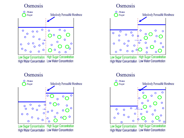
                       net movement of water molecule from higher water potential to lower water potential across partially permeable membrane


Water potential: ability of a solution to give away free water molecules. a dilute solution has higher water potential than a concentrated solution. distilled water has the higher water potential of zero. water potential of a solution is a negative value


Solute potential:  it is the ability of the solute in the solution to lower the water potential
 *a concentrated solution which has low water potential also has lower solute potential. whereas a dilute solution which has a high water potential will have a higher solute potential


higher solute potential= less negative value
lower solute potential= more negative value


For plant cells:

               Ψ=Ψs+Ψp

                                  (-)   (+)


Incipient plasmolysis: 1st stage when a plant cell starts to shrink so that there is no more pressure on the cell wall


Bulk transport~
                                     when solids or liquids pass across the cell membrane in bulk e.g, endocytosis.
                                     It is the process by which large particles pass from outside to inside the cell e.g, phagocytosis


Phagocytosis: (endocytosis)

Step 1: a bacterium attaches itself on the receptor (glycoprotein) of a phagocyte
step 2: a depression form in the plasma membrane surrounding the bacteria
step 3: a vesicle is formed called phagocytosis vesicle. a golgi vesicle buds off containing lysozymes thus forms a lysosome
step 4: this lysosome then moves toward the phagocytic vesicle and then fuses with it to form secondary vesicle
step 5: the bacteria then gets digested by hydrolytic enzymes and the debris is taken. the debris of the bacterium is taken to cell membrane through excretory vesicle


Endocytosis: the movement of solids in bulk from inside to outside the cell

*when something has to move out, it comes close to the plasma membrane through a vesicle which then fuses with the membrane passing the contents out of the cell

* Exocytosis can be through a secretory or an excretory vesicle


Pinocytosis: cell drinking
- many small vesicle carry liquid in through the plasma membrane


Friday, 5 February 2016

FOOD TESTS


Ethanol emulsion test:
- sample has to be crushed
- add alcohol to it to make a solution
- add water
- shake the tube
- if the cloudy emulsion forms then it shows presence of lipids


Starch test:
- add iodine solution to the sample 
- turns blue. black
   conclusion= starch is present 
- turns brown,
   conclusion= starch is absent 


Test for sucrose:
 sucrose is a non- reducing sugar which is made from glucose and fructose, which are reducing sugars

- performing Benedict test on a part of the sample to check for presence of reducing sugar
- if the colour stays blue, it means there are no reducing sugar present
- to check for presence of sucrose we proceed as following
1- take 2cm3 of sample
2- add 1 cm3 of HCL concentrated to it (this will on heating break down the glycosidic bonds between glucose and fructose which are reducing sugars
3- heat it and then cool it down
4- add Na2CO3 to neutralize HCL (conc)
5- add benedict solution and heat in H2O bath up to 90°C and observe the colour change


Reducing sugar test:
- add 2cm3 of sample 
- add 2cm3 of benedict reagent
- heat in a bath up to 90°C
 blue ---> red
 conclusion= reducing sugar is present 
 stays blue
 conclusion= reducing sugar is not present 


Protein test: [biuret test]
- take a sample in solution form (2cm3)
- add 2cm3 of biuret solution
- blue ---> purple
  indicates the presence of peptide bond
  conclusion= protein are present